
By Sophie Regnault with John Hutchinson and Marc Jones
Sesamoid bones are specialised, typically small, bones found in tendons near to joints, with several unusual characteristics. We’ve covered them here before. These sesamoids tend to alter the mechanics of joints, and their development also seems highly influenced by movement. They can vary between individuals or even between an animal’s left and right sides, and may even be capable of ‘regeneration’ after being cut out! For many sesamoids, it’s still not entirely clear why they’re there – why in some individuals and joints but not in others. For most people, the most familiar sesamoid bone is the kneecap (or patella), and in a previous post here I wrote about the patella (kneecap) of lizards and tuatara and what this might tell us about its evolution and function. That post is highly recommended as a primer for this one.
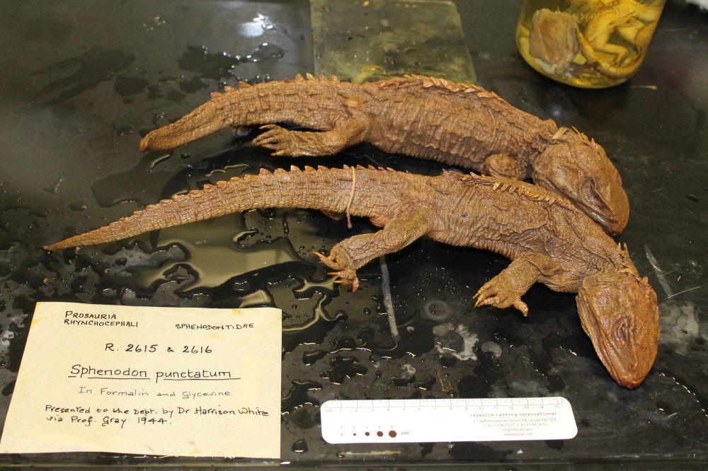
As part of the previous project on kneecaps, we scanned a bunch of preserved tuatara specimens from several different collections. Tuatara (Sphenodon punctatus) are valuable for studying evolution and anatomy. They are the closest living relatives of lizards and snakes (but separated from them by a whopping 250 million years since their most recent common ancestor) and the only species representing a once-enormous and diverse group called the Rhynchocephalia. Tuatara themselves are also rare, and for a time their survival was threatened, but with careful management populations are recovering. Outside of their native New Zealand, tuatara can be hard to obtain for study; museum specimens are often very old, valuable and difficult to replace. Their anatomy is not as well-surveyed as one might think, with characteristics like the sesamoids unknown or based off of dissections of a single specimen.

We were interested to investigate the other sesamoids in the tuatara, and what this could tell us about sesamoid evolution both in lepidosaurs (lizards + rhynchocephalians) and in general (the “big picture” in this post’s title). Evolution of sesamoid bones has been proposed to be linked to the evolution of secondary centres of ossification: the bony caps (“epiphyses”) that form on the ends of limb and other bones. If so then we would expect that (like lizards) tuatara would have many sesamoid bones throughout the body. Which ones do they possess? Did they inherit sesamoids from a common ancestor with lizards? Can the sesamoids give us clues about how sesamoids function? We took x-ray CT scans of tuatara specimens to find out.

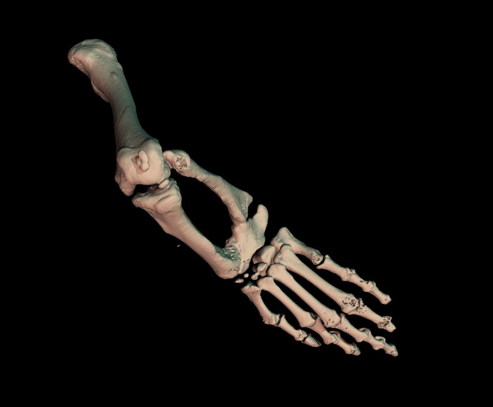
MicroCT scans of 19 tuatara showed us that they do have a similar number of sesamoid bones to lizards, but that they don’t have the same set of sesamoids. Some, like the fingertip or ankle sesamoids (AKA penultimate phalangeal sesamoids and metatarsal I-astragalocalcaneal sesamoid; see image below), are shared with lizards and likely to be inherited from a common ancestor, but others seem independently evolved in the two groups, or absent from tuatara entirely. The sequence in which the sesamoid bones mineralise during growth also seems to differ between tuatara and lizards, though not much is known about this in either group. It is likely that this sequence changes quickly throughout evolution; developmental timing can often be flexible like that.

Some sesamoids and lunulae (mineralised joint menisci; fibrous pads) were present in all tuatara, whilst others were highly variable; for example, the sesamoid bones at the fingertips (penultimate phalangeal sesamoids) were always present, but those in the palms and soles of the feet (palmar and plantar sesamoids) were only present in 77% and 27% of tuatara respectively. Amassing knowledge on sesamoids may contribute to our understanding on what sesamoids do – why they are sometimes present and sometimes not. Alone, sesamoid statistics may not tell us much, but might be useful when considered alongside other traits. For example, work by other researchers in lizards has shown that a palmar sesamoid is one of the traits associated with poorer grasping ability. This is where our future research questions are headed.

Finally, we’re very excited to be able to share the scans collected for this study, so that anybody who is interested can download, play with and research these rare and wonderful animals. The scans are available at [https://osf.io/bds35/].

Of course we are hugely indebted to the people and institutions allowing us to access these specimens: Matthew Lowe, Keturah Smithson and the University Museum of Zoology Cambridge; Susan Evans and University College London; Moya Meredith Smith and King’s College London; Steven Le Comber and Queen Mary University of London; Nicolas Di-poi and the University of Helsinki; Patrick Campbell and Natural History Museum London; Paolo Viscardi and the Horniman Museum; and all those who facilitated access and scanning. Thank you!
Read the full paper (open access) and supplementary material here.

2 Comments Add yours