Our guest post this week comes from Sophie Regnault, a PhD student in the Structure and Motion Laboratory at the Royal Veterinary College. If you would like to contribute a guest post, please get in touch on Twitter or Facebook.
The patella (kneecap) is probably the largest and most widespread of the sesamoids (a group of bones characterised by their position within tendons passing over joints), being found in mammals, birds and lizards. Lizards are thought to have evolved patellae independently from both mammals and birds. However, it’s not clear how patellae evolved and what functions they might perform in each of the three groups.
A first step in understanding patellar evolution in lizards is understanding the conditions under which the patella first appeared. Our team at the Royal Veterinary College used a statistical method called ancestral state reconstruction to trace likely patterns of gain and loss of the patella over the lizard evolutionary tree. Correlations between patellar evolution and that of other traits on the tree can provide clues about why the patella first evolved in lizards, and what benefits it might have provided then and today.
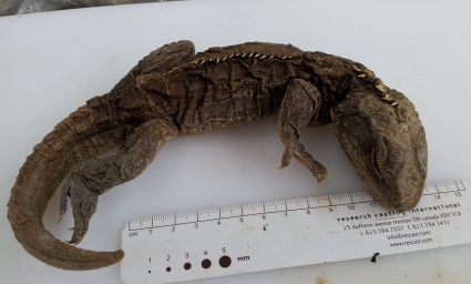
To reconstruct patellar evolution, we needed to look at both lizards and some of their near relatives for comparison (outgroups, or reference groups). One especially useful outgroup is the tuatara. This lizard-like reptile is the last survivor of a group of animals that split off from lizards 250 million years ago, and is their closest living relative. We set about studying the knee joints of tuatara to compare them with our data for true lizards.

Tuatara were thought to completely lack patellae, but after micro-CT scanning eleven adults we made the exciting discovery that four of these had some form of kneecap. The patellae looked quite different in different tuatara: some were singular and rounded, whilst others looked like several fusing pieces. This could reflect different stages of patellar development as the animals grow – even in humans, the kneecaps only develop fully around age 3. Unfortunately our tuatara had been sitting in museum pots for decades (and sometimes a century or more!), so we don’t know their ages. The lizards’ kneecaps we studied were also variable in appearance, with some single and rounded and others multipartite and possibly fusing, like the tuatara.

We also looked at one of the tuatara’s kneecaps microscopically. Unusually, the bone seems to form directly from the surrounding tendon without first laying down a cartilage template. Again, we sometimes saw the same thing in lizards, although some examples did have a cartilage template, and yet others still had mixtures of tissue types.
We aren’t sure whether the variability of the patellae in lizards and tuatara is due to different stages of development (ontogeny), or whether the kneecaps of these species are genuinely different (or even absent) in different individuals. The latter would be unusual from what we know about patellae in mammals and birds, where members of the same species all tend to have similar kneecaps (or universally lack them). However, sesamoid bones in general are notoriously variable across animal groups.

Our ancestral reconstruction isn’t clear about whether lizards and tuatara evolved kneecaps independently or whether they both inherited them from their shared ancestors. This partly results from the considerable evolutionary gap between lizards and tuatara. We looked for patellae in extinct species to try and bridge the gap, but fossils with nicely preserved knees are not very common. If present, the patellae would be very small and lack a characteristic shape, and they may also be less likely to be preserved (if calcified rather than made of bone, like in some of our lizards), or removed during the extraction of the fossil.

Despite the unclear origin of the kneecap in lizards and tuatara, our evolutionary reconstruction and anatomical similarities between the two suggest that it might have evolved in their shared ancestors, around 250 million years ago. The origin of the patella might be correlated with an increased tendency to form bone, and/or changes in the mechanics of the knee joint. The original patella might not have had much ‘use’ – perhaps it was a by-product of bone-forming ability or knee mechanics – and only later in evolutionary time did it provide a functional benefit. Through further experiments and modelling of the knee joint in lizards, tuataras and other animals, we hope to nail down what these benefits might be.
This study is part of a larger project (and the subject of my PhD!) investigating the evolution and function of the patella, or kneecap, funded by the Leverhulme Trust. You can read more about this research on lizards and tuatara here.
Glossary
Ancestral state reconstruction: Statistical analysis which uses evolutionary trees and known features to estimate the likely evolutionary history of those features.
Chondrocytes: Cells which help build cartilage.
Micro-CT: A method using a series of multiple X-ray images to reconstruct 3D models.
Ontogeny: Development through the life history of an organism, from fertilised egg to adult.
Outgroup: A species or group (a close relation to a group of interest) used for comparison to infer evolutionary relationships.
Sesamoid: A small bone found within or associated to a tendon at a joint.
2 Comments Add yours