We are back after a break! And we have a nice series of posts planned already from numerous guest writers! To kick things off, let’s get hip to bone structure. This week’s post is from Diogo M. Geraldes, PhD CEng MIMechE MEng; a biomedical engineer in London. If you would like to write for Anatomy to You, get in touch via Facebook or Twitter.
Multi-disciplinary collaborations of anatomists, clinicians, engineers and scientists have long tried to achieve a better understanding of the human skeleton’s structural and mechanical properties (and these efforts extend into all of vertebrate biology; the field of “mechanobiology”). Galileo Galilei made the first published comment on the mechanical function and shape of bone in one of his writings, when he observed that long bone dimensions were not linearly proportional to the animal’s size (Galilei 1638) (Figure 1, left). In 1803 an anatomical atlas by the physician Loder included some of the first drawings of the internal architecture of long bones (Figure 1, middle). Bourgery and Jacob improved upon the structural representation of the lower limb bones in their 1832 anatomical treatise (Figure 1, right), depicting the trabeculae (bony struts) inside the head and neck of the femur.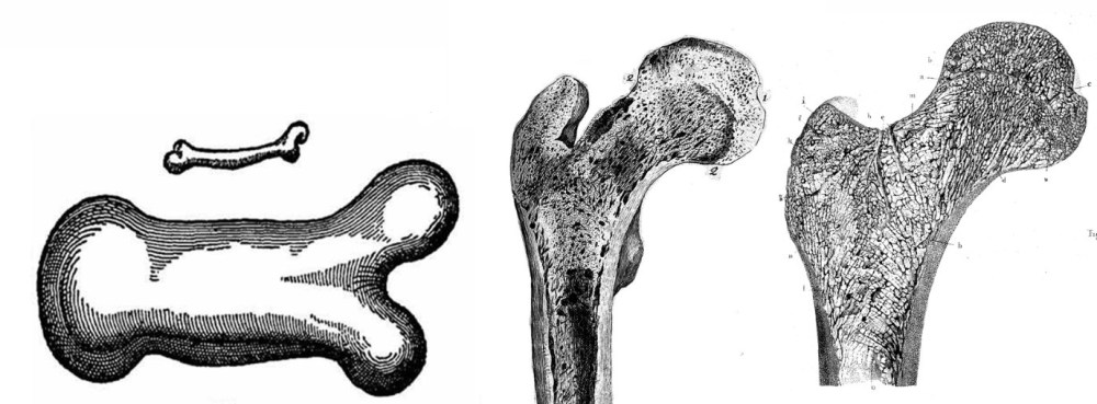
Figure 1 – Left: drawings of a small long bone and its equivalent for an animal three times larger in size (Galilei 1638); middle: drawings of the internal architecture of the proximal femur (Loder 1803)[1]; right: trabecular structures in the proximal femur (Bourgery and Jacob 1832)1; latter two images are in front-on view and are from The Wellcome Library, London under CC-BY 4.0 license.
Frederick Ward compared the main trabecular groups and a triangular area comprised of thin and loosely arranged trabeculae in the proximal (upper) femur, now called the “Ward’s triangle”, with the truss structure that supports a street lamp (Figure 2). He suggested that the alignment of the trabeculae resisted the stresses that arise from human body weight, and the shape of the femoral shaft provides the widest range of flexion and ideal moment arms (leverage) for the application of muscle forces, whilst maintaining structural strength, stability in pelvic motion and anatomical space for the thigh muscles (Ward 1838). A term sometimes used to this idea that bony struts form in alignment to best resist the forces imposed on them is the “trajectorial theory.”
The structural engineer Karl Culmann studied the drawings of the arching trabecular patterns found in different bones of the lower limb by his anatomist friend von Meyer (1867) (Figure 3, right) and noticed the coincidence of the drawn lines with the internal stress lines of similarly-shaped Fairbairn cranes undergoing analogous loading conditions (Culmann 1865) (Figure 3, left). Julius Wolff related these findings with his observations collected during his career as an orthopaedic surgeon to formulate the very famous hypothesis commonly known as Wolff’s Law (Wolff 1892):

Figure 2 – Comparison of the geometry of the proximal femur with that of a street lamp (Ward 1838); from The Wellcome Library, London under CC-BY 4.0 license.
“Every change in the form and function of the bones, or of their function alone, is followed by certain definite changes in their internal architecture, and equally definite secondary alterations of their external conformation, in accordance with mathematical laws.” (Skedros and Baucom 2007)
Figure 3 – Julius Wolff’s drawings comparing the alignment of the trabecular structures in the femur (right) with the principal stress directions of Culmann‘s analysis of a Fairbairn crane (left). Figure from “On Growth and Form” by D’Arcy Thompson; public domain.
Tobin’s work on the clinical significance of the internal architecture of the femur highlighted that femoral neck fractures often occurred in the Ward’s triangle, which becomes more distinct as bone maturity is reached, being particularly evident in women with osteoporosis (Tobin 1955). Garden’s analysis of the function of the hip joint confirmed Ward’s observations that the femoral neck is angled to promote the transmission of the loads arising from weight-bearing. He also showed that secondary trabecular groups crossed at acute angles and not at right angles, as Wolff suggested (Garden 1961), to resist the stresses induced by the complex combination of forces the femur is subjected to (Skedros and Baucom 2007).
In an attempt to use changes in the trabecular patterns as an index to evaluate the stages of deterioration of an osteoporotic bone, Singh et al. (1970) classified in detail the trabecular bone structures. Five main trabecular groups were described: the principal tensile and compressive groups, the secondary tensile and compressive groups and the greater trochanter group (Figure 4).
Figure 4 – Trabecular architecture in the proximal femur, showing the five main trabecular groups explained above. Adapted from Geraldes 2016.
The adaptation of the human skeleton’s structure to its mechanical environment has long triggered the interest of diverse scientists who have extensively analyzed and characterised the internal architecture of bones, with a focus on the proximal femur. They have established that the right-angled crossing of trabecular groups occurs in areas of bone where a single load orientation is most common. However, secondary groups crossing at other angles are required to resist the complex stresses induced by the many other frequent loading directions. This implies that bone’s structure is optimised to resist the strains and stresses arising from different daily activities; not just a simple one. Such ideas are already implemented not only in medical applications but also inspiring designs in the architecture of buildings, for example (another one here)! Who knows where these trajectories of inquiry could lead next—it is an exciting, dynamic field of integrative science.
References
Bourgery, J. B. M. and N. H. Jacob (1832). Traite Complet De L’anatomie De L’homme. Paris, France, Delaunay.
Culmann, K. (1865). Die Graphische Statik. Zurich, Switzerland, Verlag Von Meyer & Zeller.
Galilei, G. (1638). Discorsi E Dimostrazioni Matematiche: Intorno a Due Nuoue Scienze Attenenti Alla Mecanica I Movimenti Locali. Leida, Italy.
Garden, R. S. (1961). “The Structure and Function of the Proximal End of the Femur.” Journal of Bone and Joint Surgery: British Volume 43-B(3): 576-589.
Geraldes, D. M., Modenese, L. and Phillips, A. T. M. (2016). “Consideration of multiple load cases is critical in modelling orthotropic bone adaptation in the femur”. Biomechanics and Modeling in Mechanobiology 15(5): 1029-1042
Humphry, G. M. (1856). A Treatise on the Human Skeleton Cambridge, UK, Macmillan and co.
Loder, J. C. v. (1803). Tabulae Anatomicae Quas Ad Illustrandam Humani Corporis Fabricam.
Singh, M., A. R. Nagrath and P. S. Maini (1970). “Changes in Trabecular Pattern of the Upper End of the Femur as an Index of Osteoporosis.” Journal of Bone and Joint Surgery 52: 457-467.
Skedros, J. and S. Baucom (2007). “Mathematical Analysis of Trabecula ‘Trajectories’ in Apparent Trajectorial Structures: The Unfortunate Historical Emphasis on the Human Proximal Femur.” Journal of Theoretical Biology 244: 15-45.
Takechi, H. (1977). “Trabecular Architecture of the Knee Joint.” Acta Orthopaedica 48(6): 673-681.
Tobin, W. J. (1955). “The Internal Architecture of the Femur and Its Clinical Significance: The Upper End.” Journal of Bone and Joint Surgery 37(1): 57-88.
von Meyer, G. H. (1867). “Die Architektur Der Spongiosa.” Archiv für Anatomie Physiologie und wissenschaftliche Medicin 34: 615-628.
Ward, F. O. (1838). Outlines of Human Osteology, Renshaw.
Wolff, J. (1892). The Law of Bone Remodelling (Translated by Maquet, P. And Furlong, R.). Berlin, Heidelberg, New York, London, Tokyo, Springer-Verlag.

Awesome!
LikeLiked by 1 person