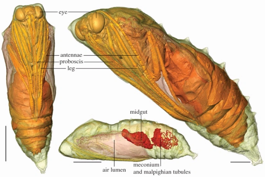
Ever wondered what biological witchcraft is going on inside the chrysalis (‘cocoon’) of a developing butterfly? One team from Manchester University and the Natural History Museum has a better idea than most – Tristan Lowe and colleagues used CT scanning technology to trace the development of living animals undergoing metamorphosis. Their use of CT ingeniously exploits the static nature of the chrysalis, and they could scan the same individuals multiple times through their transformation to make sure their results accurately represented the whole process. As the first study of its kind, this work revealed some unexpected secrets from the caterpillar, charting the development of internal organ systems as well as the dramatic changes to the external body plan. For example, they observed the rapid appearance of the tracheal (airways) system within just 12 hours of pupation. The gut, which is altered and reduced in size in adulthood due to the change in diet from leaves to nectar, begins to regress almost immediately and was found to reach its adult state by day 13. The above image comes from scans taken at 16 days, towards the end of metamorphosis, with adult features such as the eyes, legs and antennae clearly visible.
Image courtesy of Tristan Lowe and colleagues, and Royal Society publishing. See the full article here.