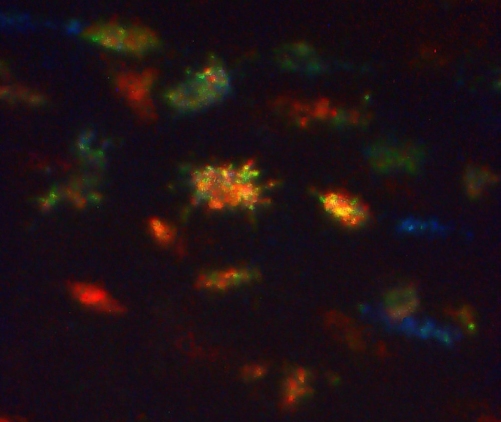
This rather abstract-looking image shows a tiny patch (around 110 micrometres across) of an ancient hominid femur. ‘Lucy’ is one of the the oldest and probably the most famous early human-like primate, belonging to the species Australopithecus afarensis. She lived around 3.2 million years ago in sub-Saharan Africa, and her remains were discovered in 1974 in Ethiopia. The forms we see here are cell spaces for osteocytes, visualised with a portable confocal microscope. The spacing and orientations of the cells can reveal crucial details about the formation of the bone, including levels of deposition and resorption during growth and the biomechanical efficacy of the bone. These results support assessments from other skeletal structures that Lucy was adapted to bipedal walking and indicate that she had a metabolic rate in line with predictions for a primate her size. This incredible technique allows researchers to study microscopic bone structures without the need for histology and other destructive methods of sampling which can damage these very precious fossils, and opens the door for further studies of the bones in these ancient skeletons.
Image courtesy of Timothy Bromage and the Ethiopia National, Addis Ababa.
Osteocytes: Bone cells, formed when osteoblasts become encased in secreted bone matrix.
