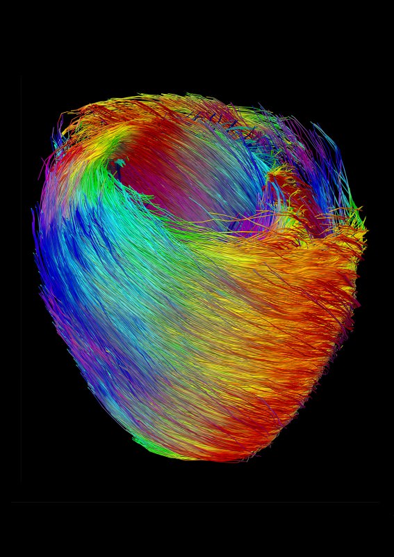
The fibres of a mouse heart resemble a neat bird’s nest structure, but their specific orientations and interactions come together to drive blood around the body. The individual muscle strands were visualised using diffusion tensor imaging, which essentially tracks the movement of water molecules through single cells, revealing their position, size and shape. The pattern of muscles in the heart is crucial. Fibres spiral out around the left ventricle, the chamber which pumps oxygenated blood out of the heart and around the body, and curve in different directions on the inside and outside of this chamber. The result, when the fibres contract, is an upwards, slightly twisting contraction of the left side of the heart, driving the blood out into the aorta at high enough pressure to continue around the body. This image demonstrates the complexity of heart muscle structure and the sheer number of fibres involved in every beat of a tiny heart.
Image courtesy of Laurence Jackson.
Aorta: The major artery which leaves the left ventricle of the heart, carrying oxygenated blood out to the body tissues.

Too glorious
LikeLiked by 1 person
Beautiful, isn’t it?
LikeLiked by 1 person