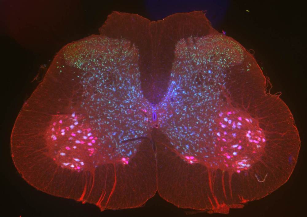
Lumbar (lower) spinal cord, sectioned and labelled using fluorescent markers. The markers attach to antibodies which are introduced by the researcher and designed to bind to specific proteins. This technique is called immunofluorescence. Here, green indicates markers for sensory neurons, red shows motor neurons and blue highlights rough endoplasmic reticulum – where most protein synthesis takes place in the cell. You can see labelled sensory axons, the long, thin part of a nerve cell, exiting the spinal cord at the bottom.
Image courtesy of Haley Titus-Mitchell. Read more about Haley’s work here.
